Home » Without Label » Abdominal Anatomy - Abdominal Exploration Series Normal Anatomy Medlineplus Medical Encyclopedia / We're going to take apart a plastic anatomy model and see what we can find in the abdomen.
Abdominal Anatomy - Abdominal Exploration Series Normal Anatomy Medlineplus Medical Encyclopedia / We're going to take apart a plastic anatomy model and see what we can find in the abdomen.
Abdominal Anatomy - Abdominal Exploration Series Normal Anatomy Medlineplus Medical Encyclopedia / We're going to take apart a plastic anatomy model and see what we can find in the abdomen.. The image also shows the pelvis, uterus, and urinary. I mean, the abs are the muscle. Overview the abdomen contains many vital organs: The rectus abdominis connects to the xiphoid process, a bony landmark at the bottom of the sternum. The regions occupied by stomach are epigastric, umbilical and hypochondriac regions.
The regions occupied by stomach are epigastric, umbilical and hypochondriac regions. The abdomen is the front part of the abdominal segment of the trunk. These two apertures, together with abdominal walls, bound the abdominal cavity. The abdomen is also known as the belly. It is the long, flat muscle that extends vertically between the pubis and the fifth, sixth, and seventh ribs.
Abdomen Wikipedia from upload.wikimedia.org You go to the gym to train your abs. The majority of these organs are encased in a protective membrane termed the peritoneum. We'll identify as many organs as we can, see how they fit into the. It is bounded superiorly by the xiphoid process and costal margins, posteriorly by the vertebral column and inferiorly by the pelvic bones and inguinal ligament. These two apertures, together with abdominal walls, bound the abdominal cavity. I mean, the abs are the muscle. Stomach is a muscular bag forming the most distensible part of the human digestive system. When you think of abs, what muscle do you typically think of?
The stomach, the small intestine (jejunum and ileum), the large intestine (colon), the liver, the spleen, the gallbladder, the pancreas, the uterus, the fallopian tubes, the ovaries, the kidneys, the ureters, the bladder, and many blood vessels (arteries and veins).
Skin, superficial fascia, muscles and associated fascia, and parietal peritoneum. This mri abdomen axial cross sectional anatomy tool is absolutely free to use. Stomach is a muscular bag forming the most distensible part of the human digestive system. The anatomy of your abdominal muscles. Explore over 6700 anatomic structures and more than 670 000 translated medical labels. Abdomen, in human anatomy, the body cavity lying between the chest or thorax above and the pelvis below and from the spine in the back to the wall of abdominal muscles in the front. But in actuality there are 4 separate muscles that contribute to your overall abdominal development. If you plan to enter a healthcare profession such as nursing, this is something you'll use on the job when performing abdominal assessments (and while documenting). The region occupied by the abdomen is called the abdominal cavity, and is enclosed by the abdominal muscles at front and to the sides, and by part of the vertebral column at the back. Together, these three turn nutrients into usable energy, as well as help dispose of solid waste. Connective tissue called the mesentery holds the abdominal organs together. The abdomen contains all the digestive organs, including the stomach, small and large intestines, pancreas, liver, and gallbladder. It is an artery, meaning that it carries blood away from the heart.
Ct, mri, radiographs, anatomic diagrams and nuclear images. We'll identify as many organs as we can, see how they fit into the. It is the long, flat muscle that extends vertically between the pubis and the fifth, sixth, and seventh ribs. You can't have a strong, muscular physique without a healthy, stable core. Abdomen, in human anatomy, the body cavity lying between the chest or thorax above and the pelvis below and from the spine in the back to the wall of abdominal muscles in the front.
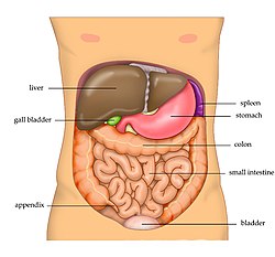
Abdominal Pain Colon Intestine Right Side Pain Real Anatomy Canstock from comps.canstockphoto.com It is the long, flat muscle that extends vertically between the pubis and the fifth, sixth, and seventh ribs. You go to the gym to train your abs. Connective tissue called the mesentery holds the abdominal organs together. The liver, stomach, and abdominal contents are clearly identified and labeled, including the cecum, ascending colon, transverse colon, descending colon, and small intestine. Overview the abdomen contains many vital organs: We'll identify as many organs as we can, see how they fit into the. The abdomen is the part of the trunk which lies just below the diaphragm. The abdomen is the part of the body that contains all of the structures between the thorax (chest) and the pelvis, and is separated from the thorax via the diaphragm.
The abdominal aorta enters the abdomen through the diaphragm at the level of the twelfth thoracic vertebre and continues to just below the umbilical area, where it splits into the right and left common iliac arteries.
We're going to take apart a plastic anatomy model and see what we can find in the abdomen. In anatomy and physiology, you'll learn how to divide the abdomen into nine different regions and four different quadrants. The area occupied by the abdomen is called the abdominal cavity. Walls of the inguinal canal. I mean, the abs are the muscle. The diaphragm is its upper boundary. Skin, superficial fascia, muscles and associated fascia, and parietal peritoneum. The abdominal wall surrounds the abdominal cavity, providing it with flexible coverage and protecting the internal organs from damage. You can't have a strong, muscular physique without a healthy, stable core. Its superior aperture faces towards the thorax, enclosed by the diaphragm. This might sound like a strange question, right? This anatomy section promotes the use of the terminologia anatomica, the international standard of anatomical nomenclature. The abdomen is the front part of the abdominal segment of the trunk.
The abdomen is the part of the trunk which lies just below the diaphragm. Overview the abdomen contains many vital organs: The area occupied by the abdomen is called the abdominal cavity. The majority of these organs are encased in a protective membrane termed the peritoneum. A point midway between the anterior superior iliac spine and the pubic symphysis.
Abdomen Wikipedia from upload.wikimedia.org Pathway by which structures can pass from the abdomen wall to the external wall. The abdomen (colloquially called the belly, tummy, midriff or stomach) is the part of the body between the thorax (chest) and pelvis, in humans and in other vertebrates. It is the long, flat muscle that extends vertically between the pubis and the fifth, sixth, and seventh ribs. This might sound like a strange question, right? The anterolateral abdominal wall consists of four main layers (external to internal): Together, these three turn nutrients into usable energy, as well as help dispose of solid waste. The liver, stomach, and abdominal contents are clearly identified and labeled, including the cecum, ascending colon, transverse colon, descending colon, and small intestine. Overview the abdomen contains many vital organs:
It is the most complete reference of human anatomy available on web, ipad, iphone and android devices.
Learn the anatomy and function of your abdominals to achieve your dream physique. Abdominal computed tomography (ct) is a type of medical imaging procedure used to diagnose and monitor internal stomach issues, like cancer, bowel obstruction, and abdominal pain. The abdomen is the part of the body that contains all of the structures between the thorax (chest) and the pelvis, and is separated from the thorax via the diaphragm. Pathway by which structures can pass from the abdomen wall to the external wall. The abdominal aorta enters the abdomen through the diaphragm at the level of the twelfth thoracic vertebre and continues to just below the umbilical area, where it splits into the right and left common iliac arteries. Lower fibers of io and transversus muscles This anatomy section promotes the use of the terminologia anatomica, the international standard of anatomical nomenclature. The liver, stomach, and abdominal contents are clearly identified and labeled, including the cecum, ascending colon, transverse colon, descending colon, and small intestine. Use the mouse scroll wheel to move the images up and down alternatively use the tiny arrows (>>) on both side of the image to move the images.>>) on both side of the image to move the images. Explore over 6700 anatomic structures and more than 670 000 translated medical labels. Walls of the inguinal canal. These two apertures, together with abdominal walls, bound the abdominal cavity. Connective tissue called the mesentery holds the abdominal organs together.